【優(yōu)品分享】Quantifoil-UltrAuFoil®
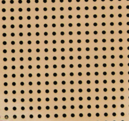
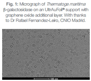
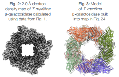
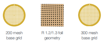
Better 3D reconstructions from less data with ultra-stable gold supports that reduce the movement of frozen specimens during imaging. During imaging at cryo-temperatures, traditional carbon supports move,particularly at the beginning of irradiation. This movement blurs images and reduces data quality. UltrAuFoil? sample supports are more conductive,and there is no differential contraction between the grid and the foil on plunge freezing. Therefore there is less crinkling of the foil during sample preparation. UltrAuFoil? R 1.2/1.3 For high-resolution transmission electron microscopy So you benefit from 1 So you benefit from Up to 0.5 ? increase in resolution on replacing traditional holey carbon supports with UltrAuFoil?.1 2 The highest resolution data from your initial movie frames before radiation damage accumulates, due to reduced beam-induced motion.2 3 3 Reduced image distortion because of lowered static and semi-mobile charge accumulation as gold is highly conductive at liquid nitrogen temperatures.2 4 Samples protected from damage due to accumulating positive charge by neutralization with secondary electrons generated by irradiating adjacent gold.2 5 Biomolecules retaining native structure and not being put under mechanical strain from foil crinkling. 6 Improved particle distribution as the gold foil is even flatter than carbon.2 7 Easier grid surveying due to the high contrast of the gold foil.2 References 1 Danev, R. et al. Routine sub-2.5 ? cryo-EM structure determination of GPCRs. Nat. Commun. 12: 4333 (2021). 2 Russo, C. R. & Passmore, L. A. Ultrastable gold substrates: Properties of a support for high-resolution electron cryomicroscopy of biological specimens. J. Struct. Biol. 193: 33-44 (2016). 3 Russo, C. R. & Passmore, L. A. Ultrastable gold substrates for electron cryomicroscopy. Science 346: 1377-1380 (2014). 4 Amil, S. M. et al & Fernandez-Leiro, R. The cryo-EM Structure of Thermotoga maritima β-Galactosidase: Quaternary Structure Guides Protein Engineering. ACS Chem. Biol. 15: 179-188 (2020). Choosing the right grid mesh and foil geometry The optimum grid and support geometry depends on many variables, but the following general guidelines will help you select the perfect UltrAuFoil? Holey Gold support. Larger particle sizes require a larger hole size to ensure sufficient numbers of particles in an image: 1 Try R 0.6/1 or R 1.2/1.3 with proteins or protein complexes. 2 For viruses and whole cells, R 2/2 is a better choice. 3 Use gold 200 mesh R 2/2 for tomography: the larger grid mesh spacing reduces the number of grid bars that might obscure tilted images. 4 R 0.6/1 is a good geometry to use with fragile complexes and membrane proteins that often need extra support. UltrAuFoil? specifications Three combinations of grid mesh size and foil geometry have been chosen to maximize compatibility with high-resolution cryo-EM data collection. Gold 300 mesh grid Grid specification: 30 squares across the grid in each direction Support geometry options: R 0.6/1 R 1.2/1.3 Ideal for: Single particle analysis Gold foil thickness: 50 nm Additional carbon layer: Please enquire for details Gold 200 mesh grid Grid specification: 20 squares across the grid in each direction Support geometry options: R 2/2 Ideal for: Cryo-electron tomography Gold foil thickness: 50 nm Additional carbon layer: Please enquire for details ecimens during imaging.


